

 脳と心理ホント
脳そのものは痛みを感じない。手術中に患者が"起きたまま"おしゃべりできる理由
 2026.10.02
 English Corner(A2〜B2・4択クイズ)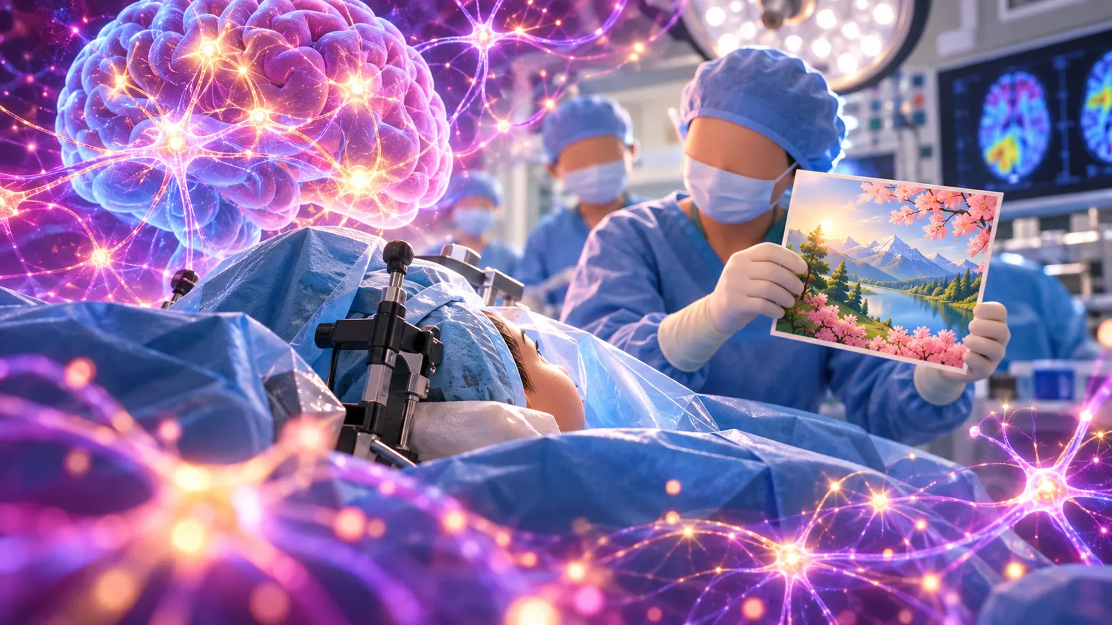
 読む前に予想してみよう!
脳の組織そのものに触れても、ほとんど痛みは感じない
痛みを感じるのは頭皮・骨膜・硬膜や太い血管など。脳の中身には痛みのセンサーがほぼない。
 研究カード(どんな研究?)
- 対象
- 神経膠腫(グリオーマ)の成人患者
- 規模
- 90報・8,091人(メタ分析)
- 研究の種類
- 観察研究のメタ分析
- 確かさ
- ★★★☆☆
脳の組織には痛みのセンサーがほぼない。だから頭皮と硬膜を麻酔すれば、起きたまま話しながら脳の手術ができる。
ひとことで言うと脳は「痛い」を感じる場所なのに、脳自体は痛くないんだって!
「痛い!」と感じているのは、脳です。
なのに、脳そのものは痛みを感じない——そう聞いたら、ちょっと混乱しませんか?
実際、脳外科には患者さんが起きたまま受ける手術があります。頭を開けたまま、医師と会話したり、絵の名前を答えたりするのです。ホラー映画のようですが、これは世界中で行われている、れっきとした治療法です。
01痛みのセンサーは「脳の外側」にあった
体のあちこちには、ケガを知らせる痛みのセンサー(侵害受容器)があります。ところが、脳の中身である神経の組織には、このセンサーがほとんどありません。
つまり「頭痛」も、脳がズキズキしているわけではありません。脳を包む膜や血管のまわりの神経が、痛みの信号を出していると考えられています。
02起きたまま手術する「覚醒下手術」
この性質を利用したのが、覚醒下手術(アウェイク・クラニオトミー)です。
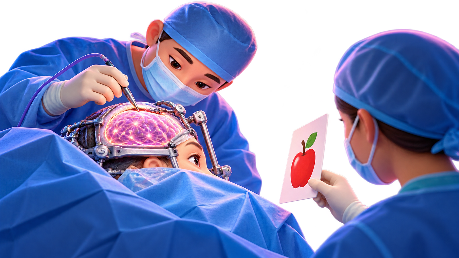
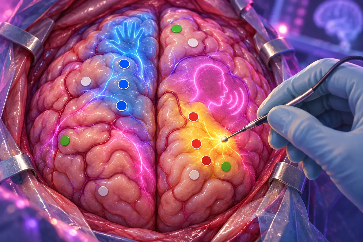
局所麻酔をかけるのは、メスを入れる頭皮や、硬膜などの「痛みを感じる部分」。施設によっては、頭を開ける間は鎮静薬で眠らせることもあります。脳の表面が見えたら患者さんに目を覚ましてもらい、医師が弱い電気で脳の一部を刺激しながら、「この絵は何ですか?」「手を握ってください」とお願いします。
もし刺激した瞬間に言葉が出なくなれば、そこは話すのに欠かせない場所。医師はそこを避けて腫瘍を取り除きます。
0390年前の「脳の地図づくり」から続く技術
この方法のルーツは、1930年代にカナダで活躍した脳外科医ペンフィールドたちの研究です。彼らはてんかんの手術中、起きている患者の脳を電気で刺激し、「手がピリピリする」「口が勝手に動く」といった反応を記録しました。こうして描かれたのが、体の各部分が脳のどこに対応するかを示す有名な「脳の地図」です。
現代の覚醒下手術も、その延長線上にあります。アメリカの1人の脳外科医が27年間に行った約860件の覚醒下手術をまとめた報告では、詳しく分析された611件のうち、マッピングが途中で中止されたのはわずか3件(0.5%)でした。
04本当に効果があるの?
神経膠腫(グリオーマ)の手術を受けた成人8,000人以上のデータを集めた2012年のメタ分析では、脳を刺激して機能の場所を確かめながら手術した場合、後に残る重い後遺症は約3.4%、確かめなかった場合は約8.2%でした。しかも腫瘍をより多く取り切れていました。
05まとめ
脳は体じゅうの痛みを感じ取る「司令塔」なのに、自分自身の痛みはほとんど感じません。その不思議な性質のおかげで、患者さんと会話しながら、言葉や動きを守る手術ができるのです。
次に頭が痛くなったら、「いま痛いのは、脳を包む膜の方かもしれない」と思い出してみてください。
この研究を英語で読もう原田英語の English CornerIt may sound strange, but the brain, which senses pain all over the body, can hardly feel pain itself. 音声
音声 4択クイズ9問A2・B1・B2タップして開く
原田英語の English Corner
この研究を英語で読もう
When you feel pain, your brain tells you. But the brain itself feels almost no pain. It has very few pain sensors. So doctors can do some brain operations while the patient is awake. They numb only the skin of the head and the thin cover of the brain.
Then they touch parts of the brain with a weak electric current. If the patient cannot speak, the doctor knows that part is important and avoids it.
76 words ・ CEFR A2
和訳を見る
痛みを感じるとき、それを教えてくれるのは脳です。しかし、脳そのものはほとんど痛みを感じません。脳には痛みのセンサーがとても少ないのです。そのため医師は、患者が起きている状態で脳の手術をすることができます。感覚をなくすのは、頭の皮膚と脳を覆う薄い膜だけです。
それから、弱い電流で脳のあちこちに触れます。もし患者が話せなくなったら、医師はその場所が大切だとわかり、そこを避けるのです。
4択リーディングクイズ
Q1. What is the passage mainly about?
脳自体はほとんど痛みを感じないので、起きたまま手術ができる、というのが全体の話題なので B。
Q2. What do doctors numb before the operation?
They numb only the skin of the head and the thin cover of the brain. とあるので D。脳の中身ではない点に注意。
Q3. In the last sentence, "avoids it" means the doctor ...
avoid は「避ける」。話すのに大切な場所なので、そこには手を出さない、という意味で A。
It may sound strange, but the brain, which senses pain all over the body, can hardly feel pain itself. In 1940, two American researchers tested which parts of the head hurt when they were touched during surgery. Pain came from the scalp, the membranes around the brain and large blood vessels, but not from the brain tissue. This fact is used in a type of operation called awake surgery.
Only the painful outer parts are numbed with local anesthesia, and the patient is woken up once the brain is exposed. The surgeon then stimulates small areas with a weak current while the patient names pictures. The surgeon avoids any spot where stimulation suddenly stops the patient from speaking, because that area is used for language, while removing the tumor.
129 words ・ CEFR B1
和訳を見る
奇妙に聞こえるかもしれませんが、体じゅうの痛みを感じ取る脳は、それ自体ではほとんど痛みを感じません。1940年、2人のアメリカ人研究者が、手術中に頭のどの部分に触れると痛むのかを調べました。痛みが生じたのは頭皮や脳を包む膜、太い血管で、脳の組織からではありませんでした。この事実は「覚醒下手術」と呼ばれる手術に利用されています。
痛みを感じる外側の部分だけを局所麻酔でまひさせ、脳が見えたところで患者に目を覚ましてもらいます。そして外科医は、患者に絵の名前を答えてもらいながら、弱い電流で小さな領域を刺激します。刺激で患者が突然話せなくなった場所は言葉に使われているので、外科医は腫瘍を取り除く際にそこを避けるのです。
4択リーディングクイズ
Q1. What did the researchers in 1940 find out?
Pain came from the scalp, the membranes ... but not from the brain tissue とあるので C。
Q2. What does the patient do during awake surgery?
The surgeon then stimulates small areas ... while the patient names pictures. とあるので A。
Q3. Why is it useful for the patient to be awake?
話せなくなった場所は言葉に必要なので避ける、と最終文にある。起きていることで重要な場所を確かめられるので D。
In an awake craniotomy, the patient stays conscious during brain surgery, and the brain itself does not hurt. Its tissue contains almost no pain receptors. In 1940, researchers stimulated different structures inside the heads of patients having surgery under local anesthesia and found that pain came from the scalp, parts of the dura and large blood vessels, not from the brain.
So once the sensitive outer layers are numbed, the patient is woken and asked to name pictures or move a hand while small areas of the cortex are stimulated with a weak current. If stimulating a spot disrupts speech, it is marked and preserved. The technique grew out of brain-mapping work by Penfield and colleagues in the 1930s.
A 2012 meta-analysis of more than 8,000 glioma patients found severe lasting deficits after about 3.4 percent of mapped operations, compared with 8.2 percent without mapping. Those figures come from observational studies, though, not randomized trials, and differences between hospitals and patients may explain part of the gap. The method does not suit every patient, but it rarely has to be stopped. One surgeon's series of about 860 cases over 27 years reported that mapping was abandoned in only 3 of the 611 cases analyzed in detail (0.5 percent).
208 words ・ CEFR B2
和訳を見る
覚醒下開頭手術では、脳の手術の間も患者は意識を保っており、脳そのものは痛みません。脳の組織には痛みの受容器がほとんどないのです。1940年、研究者たちが局所麻酔で手術を受けている患者の頭の中のさまざまな構造を刺激したところ、痛みは脳からではなく、頭皮や硬膜の一部、太い血管から生じていたのです。
そのため、痛みを感じる外側の層をまひさせたうえで、患者に目を覚ましてもらい、大脳皮質の小さな領域を弱い電流で刺激しながら、絵の名前を言ったり手を動かしたりしてもらいます。ある場所を刺激して話すことが妨げられたら、その場所には印がつけられ、温存されます。この技術は1930年代のペンフィールドらによる脳の地図づくりの研究から発展しました。
8,000人以上の神経膠腫患者を対象にした2012年のメタ分析では、後に残る重い障害が生じたのは、マッピングを行った手術で約3.4%、行わなかった手術で8.2%でした。ただし、これらの数字はランダム化比較試験ではなく観察研究によるもので、病院や患者の違いがこの差の一部を説明している可能性があります。この方法はすべての患者に適しているわけではありませんが、途中で中止しなければならないことはめったにありません。ある外科医が27年間に行った約860件の症例報告では、詳しく分析された611例のうち、マッピングを中止したのはわずか3例(0.5%)でした。
4択リーディングクイズ
Q1. What is the main purpose of the passage?
脳に痛みの受容器がないことと、それを利用した覚醒下手術の方法・効果が全体の内容なので B。
Q2. According to the 2012 meta-analysis, how often did severe lasting deficits occur when mapping was used?
severe lasting deficits after about 3.4 percent of mapped operations とあるので C。8.2%はマッピングなしの数値。
Q3. Why does the author say the results should be read carefully?
Those figures come from observational studies, though, not randomized trials, and differences between hospitals and patients may explain part of the gap とあるので D。
重要単語
- craniotomyC1名詞開頭手術The patient stayed calm during the craniotomy.
 crani-(頭蓋)+-tomy(切ること)。awake craniotomy で「覚醒下開頭手術」。
 - pain receptorB2名詞(句)痛みの受容器(センサー)The skin has many pain receptors. receptor は receive(受け取る)の仲間。receptor for ~「~の受容体」。
- anesthesiaB2名詞麻酔Local anesthesia numbs only one part of the body. local anesthesia(局所麻酔)⇔ general anesthesia(全身麻酔)の対比で覚える。英式つづりは anaesthesia。
- stimulateB2動詞刺激するThe doctor stimulated a small area with a weak current. 名詞は stimulus(刺激、複数形 stimuli)。stimulate the economy「景気を刺激する」も入試頻出。
- membraneB2名詞膜A thin membrane covers the brain. cell membrane(細胞膜)は理科系長文の定番。発音は /ˈmembreɪn/。
- mapB1動詞地図を作る、位置を特定するSurgeons map language areas before removing a tumor. 名詞「地図」だけでなく動詞で「位置を割り出す」。brain mapping「脳機能マッピング」。
- tumorB2名詞腫瘍The surgeon removed the tumor safely. 英式は tumour。remove a tumor「腫瘍を摘出する」がセット。
 入試に出る順の単語帳を印刷する →
 全5633語の「科学英単語カード」で復習する →
 動画でもっと知る
動画は各チャンネルの公式埋め込みで表示しています(YouTube)。
この話題を TED で聞く
同じテーマの TED・TED-Ed を、 TED HACK で。英語の見どころ・使える表現・学習ミッションつきの日本語ガイドです。(運営:原田英語)

 4コマでわかる
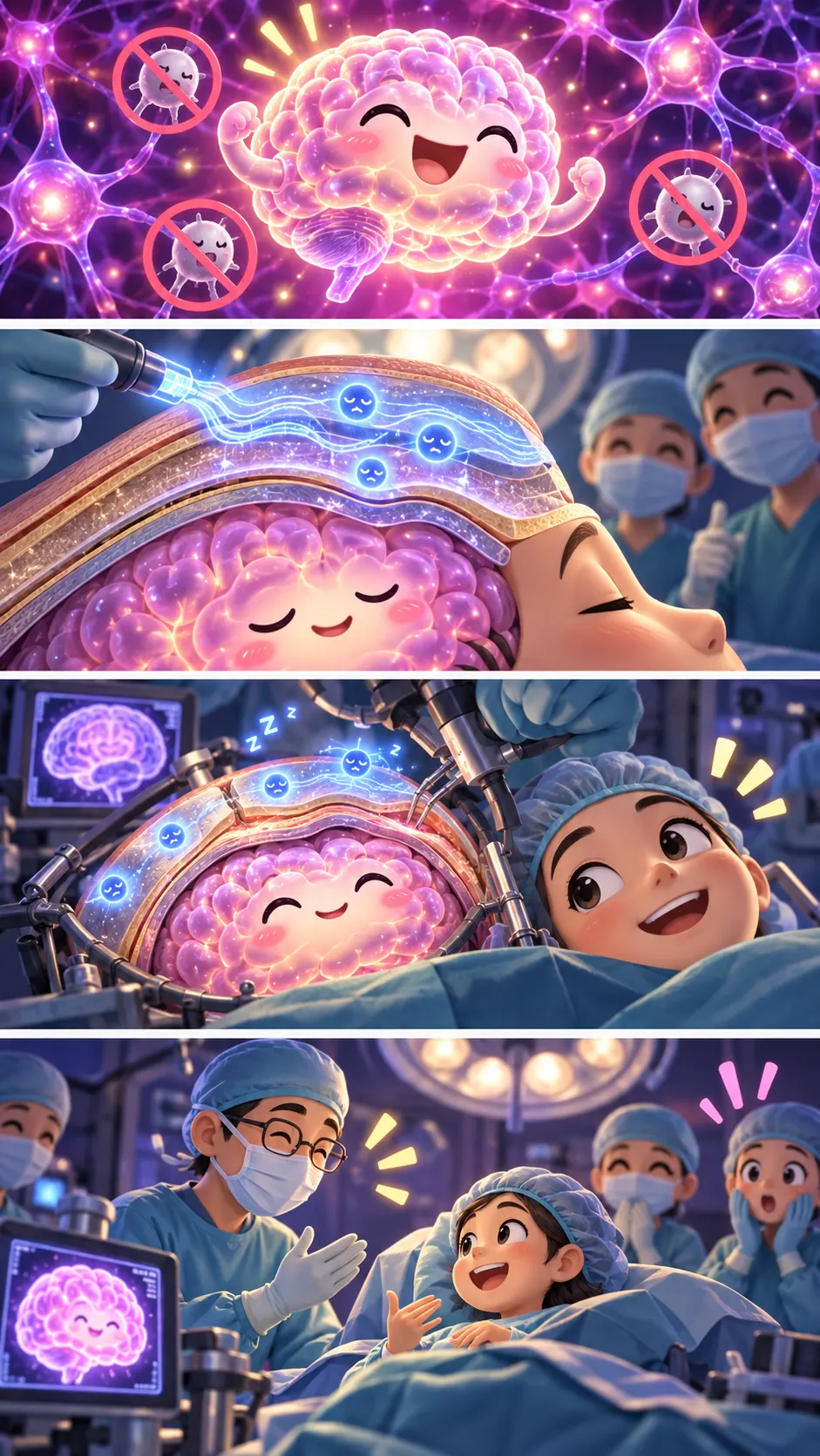
このネタ、誰かに教えたくなった?
脳の手術なのに、患者さんが起きたまま会話している…これ実際にある「覚醒下手術」。実は脳の組織そのものには痛みのセンサーがほとんどない。痛いのは頭皮や膜の方らしい。
押すと「マジかリスト」に保存されます
 出典・参考文献(4件)タップで表示
- Ray & Wolff (1940) Experimental studies on headache: pain-sensitive structures of the head and their significance in headache, Archives of Surgerydoi.org
- Penfield & Boldrey (1937) Somatic motor and sensory representation in the cerebral cortex of man as studied by electrical stimulation, Braindoi.org
- Hervey-Jumper et al. (2015) Awake craniotomy to maximize glioma resection: methods and technical nuances over a 27-year period, Journal of Neurosurgerydoi.org
- De Witt Hamer et al. (2012) Impact of intraoperative stimulation brain mapping on glioma surgery outcome: a meta-analysis, Journal of Clinical Oncologydoi.org

※本記事は上記の研究・公開資料をもとに、原田英語が独自の言葉で解説したものです。引用は著作権法第32条の範囲で行い、出典を明記しています。画像はすべてオリジナルのイメージです。研究結果には限界や個人差があります。誤りのご指摘は原田英語のお問い合わせフォームから。
 これも「マジか!?」

 脳・心理
脳・心理
脳の中の「小人」の地図が約90年ぶりに描き直された。運動の地図に"全身の司令室"が隠れていた

 脳・心理
脳・心理
絶対音感は大人になってからでも身につく? 中国や日本の音大生に多い理由と、大人の訓練実験

 脳・心理
脳・心理
赤ちゃんの頃の記憶がないのは、「記憶を作れない」からではないかもしれない


 脳・心理
脳・心理
【関学の入試に出た】地平線の月が大きく見えるのは「目の錯覚」。実は大きさは同じだった
 脳・心理
脳・心理
【どんでん返し】マシュマロを我慢できた子は成功する? 900人の追試で効果は大幅に縮んでいた

 脳・心理
脳・心理